

On 5 October 2026, the Nobel Assembly at Karolinska Institutet awarded the Nobel Prize in Physiology or Medicine jointly to Karl Deisseroth of the United States and Germany’s Peter Hegemann and Georg Nagel for discoveries concerning light-gated ion channels and optogenetics. Their work transformed a protein found in a single-celled alga into a tool that allows researchers to control selected nerve cells with light and investigate how neural circuits produce memories, emotions and behaviour.
The prize recognises a chain of discoveries that began with a basic question about how Chlamydomonas algae move towards light and eventually produced a method capable of switching defined populations of neurons on or off in living animals. The significance for neuroscience is precise rather than abstract: instead of simply observing which brain areas become active, researchers can manipulate particular cells and test what happens when those cells fire. The WP Times reports on the award as the 2026 medicine prize places optogenetics at the centre of research into the biological circuits underlying behaviour and memory. The Nobel Assembly said the method provides a way to investigate how nerve cells shape memories, feelings and behaviours in the living brain. The three laureates will share a prize of 12 million Swedish kronor.
“Optogenetics provides opportunities for mapping the brain in a way that we could once only dream of.” (Per Svenningsson, Chair of the Nobel Committee for Physiology or Medicine, Nobel Prize announcement, 5 October 2026)
The award recognises three stages of the same scientific story: Hegemann and Nagel identified the light-sensitive protein channelrhodopsin, while Deisseroth and his colleagues demonstrated how it could be used to control neurons.
How a light-sensitive algal protein became a tool for controlling neurons
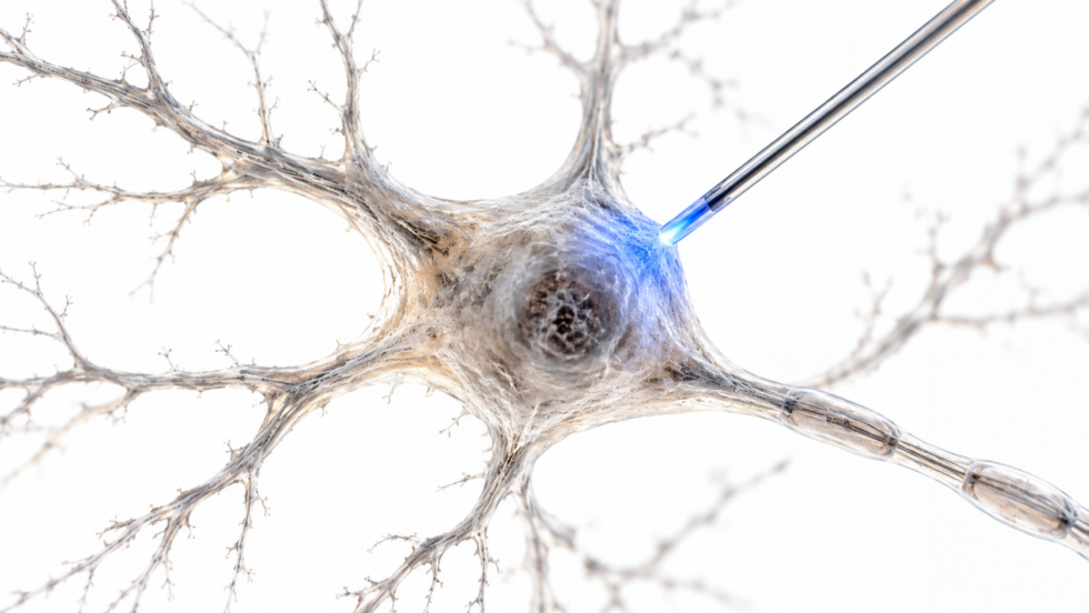
The starting point was not a search for a way to manipulate the human brain. Peter Hegemann was interested in a much simpler biological problem: how a single-celled alga detects light and moves towards a suitable source. During research in the early 2000s, Hegemann and Georg Nagel investigated proteins on the surface of Chlamydomonas and identified channelrhodopsin. The protein is a light-gated ion channel. When exposed to blue light, it opens and allows charged ions to cross the cell membrane, producing an electrical response. That mechanism immediately suggested a possibility beyond algae. If the gene encoding channelrhodopsin could be introduced into another type of electrically active cell, light might be used to control that cell in a similarly direct way.
The crucial point was that the protein did not merely detect light. It converted light into a change in electrical activity.
| Stage | Scientific development | Why it mattered |
|---|---|---|
| Algal research | Hegemann and Nagel identified channelrhodopsin | Revealed a naturally occurring light-gated ion channel |
| Neuronal experiments | Deisseroth introduced the channelrhodopsin gene into nerve cells | Made neurons responsive to light |
| Living animals | Light was delivered to genetically modified neurons in mice | Demonstrated control of neural activity in the brain |
| Optogenetics | Genetic targeting and optical stimulation were combined | Allowed specific neural circuits to be tested with high temporal precision |
The critical breakthrough was turning a biological light sensor into a controllable switch for electrically active cells.
The subsequent work established that channelrhodopsin could operate in neurons. Stanford records describe Deisseroth’s 2005 work showing that light-sensitive proteins could be expressed in mouse neurons and activated with blue light. Stanford Ingenieurwissenschaften
Stanford’s account of the technology also records the later development of optical interfaces capable of targeting neural populations in living animals with millisecond-scale precision. Deisseroth Lab
What did Karl Deisseroth change about neuroscience?
Karl Deisseroth’s contribution was to turn the molecular discovery into a practical method for neuroscience. In the 2005 breakthrough, his team introduced the gene encoding channelrhodopsin into neurons, allowing blue light to trigger electrical activity. By 2007, the approach had been demonstrated in the brains of living mice.
The difference from earlier techniques was the level of control. Electrical stimulation can activate neurons near an electrode, making it difficult to distinguish between different cell types and pathways. Optogenetics offered a way to define the cells genetically and then use light to manipulate those selected neurons. That combination of genetics and optics gave researchers two forms of precision at once. Genetic targeting determines which cells carry the light-sensitive protein, while the timing and location of illumination determine when and where those cells are activated. Stanford describes the technology as enabling researchers to interrogate defined neural circuits with millisecond-scale optical control. Its applications have since extended across studies of movement, motivation, memory, psychiatric disorders and neurodegenerative disease.
“I thought it would work but wasn't sure it would quite reach this point.” (Karl Deisseroth, Stanford University account of the development of optogenetics)
The method also evolved beyond simply switching neurons on. Researchers developed light-sensitive proteins capable of inhibiting neural activity, allowing experiments in which particular circuits could be activated or suppressed. HHMI records that engineered channelrhodopsins later provided researchers with tools for controlling neural activity in both directions. HHMI
Why controlling individual neurons matters
The brain does not operate through isolated regions that each correspond neatly to one behaviour. Neural functions emerge from networks of cells connected to one another, often with different populations performing opposing or complementary functions. A method that can selectively manipulate one population can therefore answer a different question from conventional brain imaging. Instead of asking whether an area becomes active during a behaviour, scientists can test whether changing the activity of a defined group of neurons alters that behaviour.
This distinction is central to the importance of optogenetics. It turns correlation into an experimental test of causation within neural circuits.
How optogenetics helps scientists study memories
Memory research provides one of the clearest examples of what the technology makes possible. Researchers have used optogenetics to identify populations of neurons associated with particular experiences and then manipulate those cells to investigate their contribution to memory formation and recall. Experiments in mice have shown that specific groups of neurons can become associated with a particular experience. When those cells are subsequently reactivated with light, researchers can investigate whether elements of the original memory or behavioural response are reproduced.
HHMI has documented experiments in which researchers used optogenetics to identify neurons associated with a particular setting and later reactivate that selected population with light. The work demonstrated how the method can be used to investigate the cellular basis of memory rather than treating memory as an activity spread uniformly across an entire brain region. The Nobel Committee’s description places these findings within a wider field of research into memories, feelings and behaviour. The method does not mean that a complete human memory can simply be switched on or off. Rather, it gives researchers an experimental way to identify and manipulate components of the neural circuits involved. Several areas of research have benefited from this approach:
- memory formation and recall;
- emotional responses and fear-related behaviour;
- motivation and reward;
- movement and motor control;
- mechanisms associated with neurological disease;
- neural circuits implicated in psychiatric disorders.
The important advance is the ability to manipulate selected neurons while leaving neighbouring populations comparatively untouched.
That precision has helped researchers investigate why apparently similar brain regions can produce very different effects depending on which cells within them are activated.
Why the Nobel Prize focuses on channelrhodopsin
The Nobel award is formally for discoveries concerning “light-gated ion channels and optogenetics”. Channelrhodopsin is central because it supplied the molecular mechanism that made optical control of electrical cells possible. The protein is found naturally in algae, where it forms part of the organism’s light-sensing system. When blue light reaches it, the channel opens and positively charged ions enter the cell. In a neuron engineered to express the protein, the same basic process can trigger an action potential. That is a striking example of a mechanism moving between biological systems. A protein that evolved to help a microscopic organism respond to its environment became a component of an experimental system for investigating mammalian brains. The researchers did not need to create the fundamental light-sensitive mechanism from scratch. They identified it in nature, established how it worked and adapted it for another cellular context.
“The laureates have laid the foundation of a new era in neuroscience.” (The Nobel Assembly at Karolinska Institutet, Nobel Prize in Physiology or Medicine announcement, 5 October 2026)
The technology has continued to develop since the first demonstrations. Stanford researchers now work with channelrhodopsins engineered for different wavelengths, speeds and functions, including systems designed to inhibit rather than stimulate neurons.
What could optogenetics mean for medical treatment
Optogenetics remains primarily a research technology, but its potential clinical applications are one reason the field has attracted sustained attention. The Nobel Committee specifically notes research aimed at restoring sight in people with visual impairment. The principle is to make selected cells responsive to light and then deliver controlled illumination to them. In experimental medicine, this approach can be considered where conventional electrical or pharmacological methods cannot provide the required cellular specificity.
Research into optogenetics has also been connected with neurological and psychiatric conditions. Scientists use the technique to investigate circuits involved in disorders rather than assuming that symptoms originate from an entire brain region. The distinction matters because conditions such as depression, addiction, Parkinson’s disease and other neurological disorders involve complex networks. Stanford describes optogenetics as a platform used to investigate neural circuitry relevant to neurodegenerative disease, mood disorders and potential therapeutic development. At present, the most established role of optogenetics is therefore experimental rather than routine clinical care. Its value lies in allowing researchers to establish which cells and circuits matter before considering whether similar mechanisms could eventually be translated into treatments.
From laboratory mice to clinical research
The move from animal experiments to human medicine presents additional technical and safety requirements. Delivering genes to precisely defined cells, reaching those cells with light and maintaining control without damaging tissue are separate engineering and medical challenges. The technology itself, however, has already demonstrated that neural activity can be manipulated with far greater specificity than was possible with many earlier approaches. The Nobel Committee says researchers are using optogenetic methods in attempts to restore sight in people with visual impairment. That places the technique beyond a purely conceptual neuroscience tool and into an area where optical and genetic engineering intersect with clinical medicine.
Who are the three 2026 Nobel medicine laureates
Karl Deisseroth, Peter Hegemann and Georg Nagel come from different scientific backgrounds, but their work converged around the same biological mechanism. Karl Deisseroth, born in 1971, is a professor of bioengineering and psychiatry and behavioural sciences at Stanford University and an investigator with the Howard Hughes Medical Institute. Stanford lists his PhD in neuroscience from 1998 and MD from 2000.
Peter Hegemann, born in 1954, is a Hertie Senior Professor of Neuroscience at Humboldt University of Berlin. The Nobel Assembly attributes the prize-winning discoveries to work carried out at the Max Planck Institute for Biochemistry. Georg Nagel, born in 1953, is a professor of molecular plant physiology at the University of Würzburg. His prize-winning work was conducted at the Max Planck Institute for Biophysics in Frankfurt, according to the Nobel Assembly. Their contributions can be separated broadly into three stages:
- Hegemann investigated how algae respond to light and identified the molecular machinery behind that response.
- Nagel worked with Hegemann on the characterisation of channelrhodopsins and their light-gated ion-channel properties.
- Deisseroth adapted channelrhodopsin for use in neurons and demonstrated optical control of neural activity in living animals.
The three strands turned an observation in a microscopic organism into a method now used across neuroscience laboratories worldwide.
What happens next for optogenetics research
The 2026 Nobel Prize recognises a method that is already deeply embedded in neuroscience rather than a technology waiting for its first application. Current research is focused on improving the precision, depth, wavelength and reliability of optical control while expanding the range of cells that can be targeted. One direction involves developing proteins that respond to longer wavelengths of light, which can penetrate tissue more effectively. Another involves engineering inhibitory tools so researchers can suppress rather than activate defined neurons. Stanford’s technology programme lists red-shifted channelrhodopsins, inhibitory variants and systems for more selective genetic expression among continuing areas of development.
The wider scientific question remains how patterns of activity across enormous numbers of interconnected cells produce memory, emotion and behaviour. Optogenetics does not answer that question by itself, but it gives researchers a way to intervene in individual components of those networks and measure what follows.
For the three 2026 laureates, the path began with a light-sensitive protein in Chlamydomonas. It ended with a method that lets neuroscientists use light as an experimental switch inside living brains.
Materials used: Nobel Prize, Nobel Assembly at Karolinska Institutet, Stanford University, Stanford School of Engineering, Stanford Deisseroth Lab, Howard Hughes Medical Institute.
Read about the life of Westminster and Pimlico district, London and the world. 24/7 news with fresh and useful updates on culture, business, technology and city life: When will Oasis 2027 tour dates be announced and could 12 Etihad nights define Manchester next summer